Paget's disease of bone is a chronic disease of the skeleton. It is a common disease in older people, occurring in about 3 percent to 4 percent of the population over age 50. It is slightly more common in men than women.
In normal bone, a process called remodeling takes place every day. Bone is absorbed and then reformed in response to the normal stresses on the skeleton.
- Cells of the bone called "osteoclasts" absorb bone.
- Cells of the bone called "osteoblasts" make new bone.
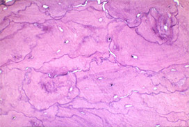
In Paget's disease, osteoclasts are more active than osteoblasts (Figure 1). This means there is more bone absorption than normal. The osteoblasts try to keep up by making new bone, but they overreact and make excess bone that is very chaotic (Figure 2). The new bone is abnormally large, is deformed and fits together haphazardly. Normal bone has a tight overlapping structure, like a well-constructed brick wall. Bone afflicted by Paget's disease has an irregular mosaic pattern, as though the bricks were just dumped in place. The end result is bones that are large and dense, but weak and brittle. The bone is prone to fractures, bowing and deformities.
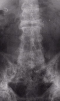
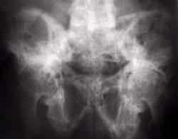
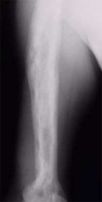
Paget's disease can affect any bone in the skeleton. It appears most often in the spine (Figure 3), pelvis (Figure 4), long bones of the limbs (Figure 5) and skull. It can be present in just one bone or in several bones. It can affect the entire bone or just part of it.
A doctor can usually diagnose Paget's disease by looking at an X-ray. An affected bone appears larger and more dense than usual. It can have a deformed shape. In the very early stages of Paget's disease, when there is just overactive absorption going on, it can look like there is a hole in the bone. Later in the disease process, this darker area can take the shape of a "V," with denser, thicker bone following behind it.
A blood test called serum alkaline phosphatase can also be used by a doctor to confirm the diagnosis. This test measures bone turnover. It is usually quite elevated in patients with Paget's disease. A doctor can also detect Paget's disease with urine tests that show rapid bone turnover.A doctor can usually diagnose Paget's disease by looking at an X-ray. An affected bone appears larger and more dense than usual. It can have a deformed shape. In the very early stages of Paget's disease, when there is just overactive absorption going on, it can look like there is a hole in the bone. Later in the disease process, this darker area can take the shape of a "V," with denser, thicker bone following behind it.
A doctor may use bone scans to see if more than one bone is involved. In this test, a radioactive material is injected into a vein to travel throughout the body. A special camera is used to look through the whole skeleton for "hot spots," areas where there is more bone turnover than usual. Paget's disease almost always looks "hot" on a bone scan, except when it has been there for a long time and has "burned out."
Biopsy of the bone is sometimes necessary to confirm the diagnosis of Paget's disease, to be sure it is not something else, and sometimes before the doctors can give medication to help treat symptoms. These biopsies are sometimes done with a needle, using numbing medicine only, or with a little sedation. Sometimes they are done in the operating room through small incisions.
Risk Factors/Prevention
Paget's disease tends to appear in families. It can be present in as many as 25 percent to 40 percent of the relatives of someone with the disease. It is also more common in people of Anglo-Saxon descent and those who live in certain geographic areas, such as England, the United States, Australia, New Zealand and Western Europe. It is not common in Scandinavia, China, Japan or India.Some doctors suggest that some environmental exposure, not just genetics, is important in the development of Paget's disease. This has not been proven definitively.
Paget's disease is uncommon in people younger than 40 years old. It is more common as people age. There are no known ways to prevent Paget's disease from occurring. Eating a healthy diet with sufficient calcium and vitamin D and getting exercise are important components in maintaining skeletal health and joint mobility.
Symptoms
People with Paget's disease often do not have any symptoms at all. A doctor may notice Paget's disease on an X-ray that is taken for another reason or during routine blood work when an elevated serum alkaline phosphatase level is found.In patients who do have symptoms, bone pain is the most common complaint. This pain can be related to active Paget's disease or its complications, which include:
- Fractures of brittle bone
- Deformity of bone
- Advanced arthritis of joints near affected bone
- Compression on neighboring nerves from enlarged bones, leading to a loss of sensation or movement
Symptoms can arise from the effect on calcium levels in the blood stream. When Paget's disease is active in several bones, the overactive osteoclasts can release enough calcium from the bone as they break it down to cause an elevated calcium level in the blood. This rare complication might cause fatigue, weakness, loss of appetite, abdominal pain, or constipation. A doctor can also detect Paget's disease from its effect on the heart if excess blood supply is shunted to overactive bones in severe cases.
When pain is severe and unrelenting in an area affected by Paget's disease, the disease may have degenerated into a bone cancer. Paget's sarcoma (Figure 6) occurs in only about 1 percent of patients with Paget's disease. These patients are usually older than 70 years of age. This type of malignant bone tumor is very aggressive and carries a poor prognosis. None of the medical treatments for Paget's disease have had an effect on the risk for the development of Paget's sarcoma.
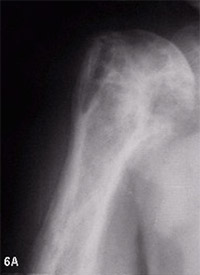
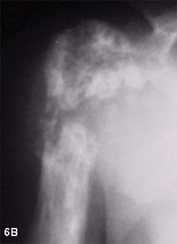
Figure 6: Paget's sarcoma of the humerus. There has been a significant change over time in the X-ray appearance from 6A to 6B. There is actual bone destruction in the later X-ray (6B) and there was a large soft tissue mass outside of the bone on the MRI.
Non-surgical Treatment Options
Patients with no symptoms require no treatment besides observation. Periodic X-rays may be recommended so the doctor can watch for any changes. For patients who have mild pain from the disease or from arthritis associated with it, nonsteroidal anti-inflammatories (NSAIDs) and aspirin can be very helpful. When the pelvis or leg is involved, using a cane can effectively decrease pain by decreasing the forces going through the bone. A cane can also help you prevent falls, so there is less risk of fracture. Braces may be used to prevent malalignment of the bones.
When bone pain is more significant, medications called bisphosphonates are the treatment of choice. These drugs block osteoclasts and can be very effective in treating Paget's disease. There are several types of bisphosphonates that are taken by mouth (oral form) or by injection (intravenous form). Your doctor will tell you which type is best for you and how long you will need to take it. Your alkaline phosphatase level should be checked periodically while you take these medications; it will steadily decline along with the bone pain. Medical treatments can help decrease the symptoms of Paget's disease, but there is no known way to reverse the effects on the bone.
Surgical Treatment Options
Surgery is used mainly to treat the complications of Paget's disease. You may need surgery if you have a fracture, if your bone is badly aligned, or if severe arthritis develops. You may also need surgery if the enlarged bone begins to compress nerves, especially in the spine or skull. In the rare cases of Paget's sarcoma, surgery is almost always used to try to remove the tumor entirely. Chemotherapy and radiation therapy may also be used.
The surgical procedures used to treat fractures, malalignment or arthritis in patients with Paget's disease are similar to those used to treat similar conditions in people with normal bone. However, because Paget's disease increases the blood supply to bones, your physician may recommend taking bisphosphonates before the surgery to reduce potential blood loss. Bones affected by Paget's disease may take longer to heal than normal bones. Longer rehabilitation than usual may be necessary.
Research on the Horizon/What's New?
Scientists are currently investigating the genes that may be involved in Paget's disease. This work could enable doctors to predict who may be at risk for the disease and lead to new therapies to treat it. The more details doctors can uncover about why the disease occurs, the better able they will be to develop treatments specific to Paget's disease. Eventually, doctors hope to reverse the effects on the bone, instead of just slowing them.
Reproduced with permission Fischer S., (interim ed): Your Orthopaedic Connection. Rosemont, Illinois. Copyright American Academy of Orthopaedic Surgeons.