CCAM Microscopy Facility

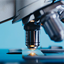
The CCAM Microscopy Facility provides the UConn Health research community as well as other academic and industrial institutes, access to its state-of-the-art equipment for quantitative fluorescence imaging applications. Within the facility, there are two laser scanning confocal microscopes equipped with 34-channel high-efficiency QUASAR detectors. One of these microscopes supports nonlinear optical (also known as 2-photon) excitation, fluorescence correlation spectroscopy (FCS), and fluorescence lifetime imaging microscopy (FLIM). Additionally, there is a light sheet microscope available for micro/mesoscale 3D imaging. Furthermore, the facility recently acquired a state-of-the-art Zeiss Elyra 7 microscope that is capable of total internal reflection fluorescence microscopy (TIRFM) and supports several super-resolution techniques such as lattice structured illumination microscopy (SIM), stochastic optical reconstruction microscopy (STORM), 3D photoactivated localization microscopy (PALM), and DNA points accumulation for imaging in nanoscale topography (DNA-PAINT).

The equipment may be used independently by authorized users who have completed the required training for each piece of equipment they choose to use. Alternatively, CCAM staff are available for assisted use. CCAM offers data storage space to each lab with an active account. Please contact Dr. Yi Wu, yiwu@uchc.edu, or Susan Staurovsky, staurovsky@uchc.edu, for additional information or assistance.

CORE Marketplace Logo

SIM of MAP2 Neuronal Culture

SIM Image of Primary Neuronal Culture, K. Caro.

LightSheet Midgut Drosophila Melanogaster cmyk colors

LightSheet Midgut Drosophila Melanogaster, K. Kannan.

SIM Image of Mitochondria

SIM Image of Mitochondria, Y. Wu.

Equipment

Microscopes and user computers are available at two different locations in Farmington, Connecticut, the Academic Research Building at the UConn Health main campus and the Cell & Genome Sciences Building. Please visit Microscopy Equipment to obtain detailed information about the instrumentation and their locations.

Fees

CCAM utilizes CORES (internal login) and CORES (external login) for all billing and scheduling events. You must be authorized by the system and have the permission to charge to an FRS account. Please check with your departmental administration to get authorization within the CORES system. Off-campus users should contact CCAM staff for billing procedures. Please see Microscopy Fees for a detailed listing of user fees.

Microscope Access

Users can opt to either be trained and use the microscopes independently or they can use the microscopes with the assistance of a CCAM staff member, via service work. Once you are trained to use the microscope independently, you may sign up for the scope during work hours or for after work hours / weekend use. Please note that you must be trained for each microscope separately. Users that opt to use the microscope via service work will be charged a different rate than the trained, independent users. Please see the Training page and Fees page for more information.

For your convenience, please use the following document for a detailed listing of the equipment and software resources available at the CCAM Microscopy Facility.

Blocks with Grant Support