Zeiss LSM 880
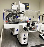
- Mounted on an Axio Observer Z1 with automated XYZ control
- Spectral 32-channel QUASAR GaAsp detector, detection range 410 – 696 nm
- Two PMT’s, detection range 415 – 735 nm, and a transmitted light detector
- Incubation system with temperature, humidity and carbon dioxide control.
| Laser Wavelengths (nm) | Type | Common Fluorophores |
|---|---|---|
| 405 nm | Diode | Alexa405, DAPI, Hoechst |
| 458, 488, and 514 nm | Argon | Alexa488, GFP |
| 561 nm | Diode Pumped Solid State (DPSS) | Alexa568, Rhodamine |
| 633 nm | Helium Neon (HeNe) | Alexa 647, Cy5 |
| Objectives | Magnification | Medium | Working Distance (mm) |
|---|---|---|---|
| EC Plan-Neofluar | 10x/0.3 | Air | 5.2 |
| Plan-Apochromat | 20x/0.8 | Air | 0.55 |
| LD LCI Plan-Apochromat | 25x/0.8 | Water, Silicone, Glycerine, Oil | 0.57 |
| Achrostigmat | 40x/1.3 | Oil | 0.21 |
| C-Apochromat | 40x/1.2 | Water | 0.28 |
| Plan-Apochromat | 63x/1.4 | Oil | 0.13 |
Zeiss Observer Z1 – TIRF
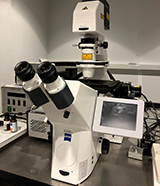
The Zeiss Observer Z1 is a motorized, inverted microscope fitted with a stage-top incubation system for live cell imaging, providing heating, humidification, and carbon dioxide. The system is equipped with a 5.5-megapixel, Andor Neo sCMOS camera. The scope is capable of phase, DIC, and fluorescence microscopy. The Lumencor Spectra 7, 6 LED light source is currently configured for imaging DAPI, GFP, Rhodamine, and Cy5. Objective choices range from 10x through 63x.
An advanced imaging feature of this system is its capability to perform TIRF using 488 nm, 514 nm, and 561 nm wavelengths for fluorescent indicators such as GFP, YFP, and RFP using the 100x/1.45 alpha-Plan Fluar oil immersion objective.
The microscope is controlled via MicroManager, a plugin from FIJI, enabling it to acquire single-channel or multi-channel images as well as time series with multiple position acquisition.
Imaging01
 This CCAM user computer is a Windows 7 computer with MetaMorph, Photoshop, Office, Zeiss LSM and Zen Image Browsers. All the software is available, with prior approval, via remote access.
Zeiss Elyra 7 SIM2 – R1521
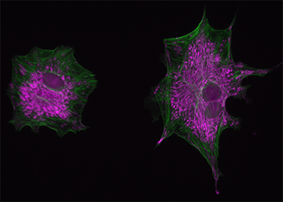
Structured illumination microscopy, SIM, is a widefield technique that greatly increases the diffraction-limited resolution of the light microscope, ~ 200 nm, to super-resolution, ~ 100 nm. SIM uses a periodic grating that is projected onto the image plane of the microscope. Software is then used to analyze the resulting Moiré pattern, and through deconvolution, a super-resolution image is obtained. The Zeiss Elyra 7 SIM2 uses an advanced reconstruction algorithm that improves the SIM resolution to less than 100 nm, along with quick image acquisition and compatibility for live-cell applications.
- Zeiss Axio Observer 7, with definite focus
- XY and Z piezo-controlled stage and focus
- Two CMOS cameras for dual acquisition
- Incubation system for temperature, humidity, and carbon dioxide
- TIRF, PALM, and STORM.
- Samples live and fixed, up to ~ 150 µm thickness
| Laser Wavelengths (nm) | Common Fluorophores |
|---|---|
| 405 nm for PALM | Alexa405, DAPI, Hoechst |
| 488 nm | Alexa488, GFP |
| 561 nm (500mW) | Alexa568, Rhodamine |
| 640 nm (500mW) | Alexa 647, Cy5 |
| Objectives | Magnification | Medium | Working Distance (mm) |
|---|---|---|---|
| EC Plan-Neofluar | 10x/0.3 | Air | 5.2 |
| Plan-Apochromat | 20x/0.8 | Air | 0.55 |
| Plan-Apochromat | 40x/1.4 | Oil | 0.13 |
| Plan-Apochromat | 63x/1.4 | Oil | 0.19 |
| C-Apochromat Corr | 63x/1.2 | Water | 0.17 |
| alpha Plan-Apochromat Corr | 63x/1.46 | Oil | 0.10 |
| Plan-Apochromat | 100x/1.57 | Oil-HI | 0.12 |
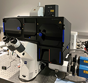
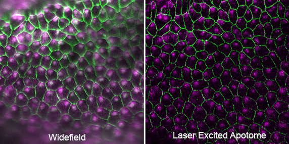
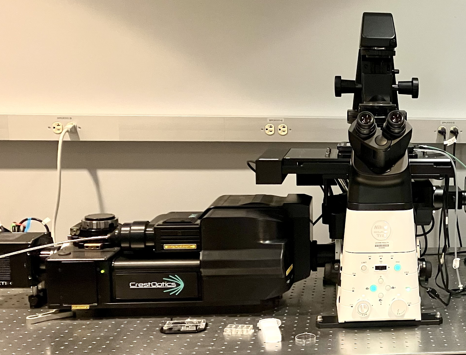
Nikon Crest V3 Spinning Disk Confocal – R1522
- Confocal and Widefield Modes for samples not exceeding 50 um
- X-Light V3 Spinning Disk by CrestOptics
- 70 um pinhole size, 250 um spacing
- Fast Imaging, and Tiling Capabilities, with Large Field of View, 25mm
- Nikon Eclipse Ti2 with Perfect Focus System, and XY Motorized Stage
- Teledyne Photometrics Kinetix 10MP sCMOS Camera
- Tokai Hit Stage Top Incubation System with CO2 and O2
Waveform Analysis of Chlamydomonas Using High Speed Acquisition Settings
| Lasers | Emission Filters | Common Fluorophores |
|---|---|---|
| 405 nm | 438/24 (426 – 450) | DAPI |
| 440 nm | 485/20 (475 – 495) | CFP |
| 477 nm | 511/20 (501 – 521) | GFP, Alexa 488 |
| 518 nm | 560/25 (548 – 572) | YFP |
| 545 nm | 595/31 (580 – 610) | CY3, Alexa 555 |
| 639 nm | 685/40 (665 – 705) | CY5, Alexa 647 |
| 745 nm | 794/32 (778 – 810) | CY7, Alexa 750 |
| Objectives | Magnification | Medium | Working Distance (mm) |
|---|---|---|---|
| Plan-Apochromat λ D | 10x/0.45 | Air | 4.0 |
| Plan-Apochromat λ D | 20x/0.8 | Air | 0.8 |
| Plan-Apochromat λ S CORR | 40x/1.25 | Silicone | 0.3 |
| Plan-Apochromat λ D | 60x/1.42 | Oil | 0.15 |
| Plan-Apochromat Ph3 | 60x/1.40 | Oil | 0.16 – 0.18 |
| Plan-Apochromat λ D | 100x/1.45 | Oil | 0.13 |
Zeiss 780 – R1523

-
- confocal/FCS/NLO system
- Axio Observer Z1.
- Spectral 32-channel QUASAR GaAsp Detector allowing for spectral imaging, maximum sensitivity photon counting, and Fluorescence Correlation Spectroscopy (FCS) as well as two adjacent PMTs.
- GaAsp detector with range of 410 – 696 nm
- Two PMTs with a range from 415 – 735 nm.
- Incubation system for temperature, humidity and carbon dioxide control.
- Becker-Hinkl Fluorescence Lifetime Imaging Microscopy (FLIM) detector on a non-de-scanned detection port, particularly useful for detection of fluorescence resonance energy transfer (FRET) based probes.
| Lasers | Type | Common Fluorophores |
|---|---|---|
| 405 nm | Diode | Alexa405, DAPI, Hoechst |
| 440 nm | Diode | LSSmOrange, Sapphire |
| 458, 488, and 514 nm | Argon | CFP, Alexa 488, YFP |
| 561 nm | Diode Pumped Solid State (DPSS) | Alexa 555, Cy3 |
| 633 nm | Helium Neon (HeNe) | Alexa 647, Cy5 |
| 720 – 950 nm | Coherent Chameleon tunable, software controlled, mode locked Ti:Sapphire laser for multi-photon excitation (NLO) | DAPI 740 nm, GFP 850 nm |
| Objectives | Magnification | Medium | Working Distance (mm) |
|---|---|---|---|
| C-Apochromat | 10x/0.45 W | Water | 1.8 |
| Plan-Apochromat | 20x/0.8 | Air | .55 |
| C-Apochromat | 40x/1.2 W | Water | .41 |
| Plan-Neofluar | 40x/1.3 | Oil | .21 |
| Plan-Apochromat | 63x/1.4 | Oil | .19 |
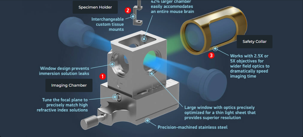
Zeiss LightSheet – R1515
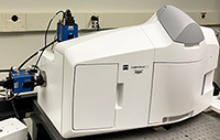 The Zeiss LightSheet is an advanced fluorescent microscope that allows you to image optical sections of large, live or fixed samples with virtually no bleaching or phototoxicity to the specimen. The optics are equipped for water-based or cleared specimens and dual sCMOS, cooled cameras to provide high sensitivity, speed, 3-D imaging of live or fixed thick specimens. The extremely fast acquisition speeds allow you to capture large amounts of data within a fraction of the time compared to other imaging techniques.
| Lasers | Emission Objectives | Excitation Objectives |
|---|---|---|
| 405 nm Diode | 2.5x/0.12 Fluar | 5x/0.1 |
| 488 nm Diode | 5x/0.16 EC Plan-Neofluar | 5x/0.1 |
| 561 nm Diode | 20x/1.0 Plan-Neofluar Clearing | 10x/0.2 |
| 633 nm Diode | 20x/1.0 Plan-Apochromat Water | 10x/0.2 |
The Translucene Biosystems imaging chamber, along with the 2.5x objective, allows for larger samples, and an ease for mounting samples. This chamber is also compatible with the 5x objective and can be used for imaging samples in higher refractive index buffers.
Zeiss AxioVert 200M – R1522
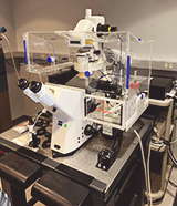 The Zeiss AxioVert 200M is a motorized, inverted microscope fitted with an incubation system for live cell imaging, providing heating, humidification, and carbon dioxide environmental conditions. The system is equipped with a pco.edge 4.2 sCMOS camera, with 95% quantum efficiency at 580nm, and 2048 (H) x 2048 (V) pixels. The maximum from rate is 40 fps at full resolution. The scope is capable of phase and DIC microscopy and is fitted with a Lumencor Spectra 7, 6 LED light source for imaging with CFP, GFP, OFP/mCherry, and Cy5. MetaMorph software is used for the acquisition and analysis of data, including the ability to capture long-term, time series with multiple positions.
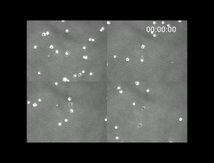
(When clicked, the video will open a new window. Duration: 43 seconds, Size: 11 MB. )
3 Day experiment, captured on this setup, observing 3T3 cells, in four different positions, was visualized using transmitted light and maintained with 5% carbon dioxide and 37-degree environmental conditions.
Imaris – R1510
 Imaris is a 64 bit computer with ImarisSurpass 3-D rendering and analysis software, MetaMorph, Photoshop, and Zeiss Zen Image Browser.
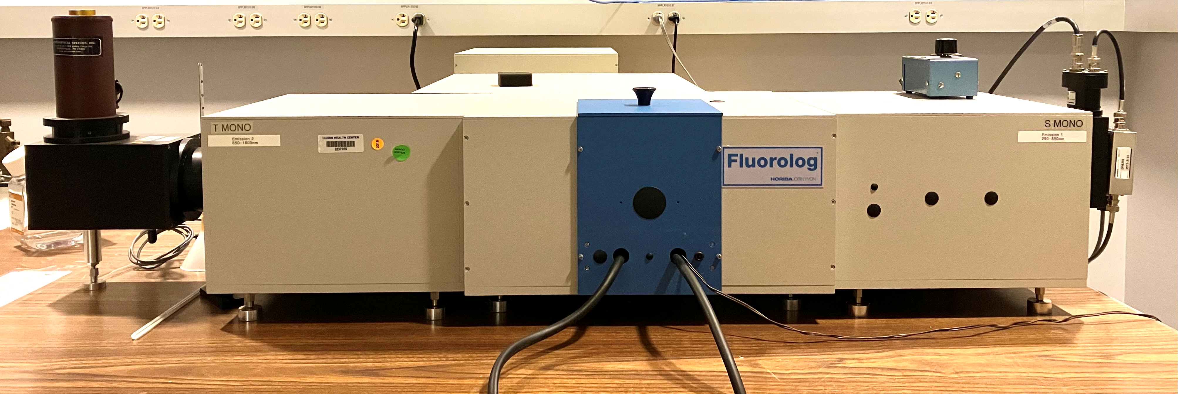
Fluorolog – R1513
CCAM Wet Lab – R1517
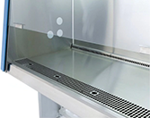 Provides tissue culture hood and tissue culture microscope, incubators, refrigeration, and bench space.