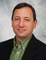
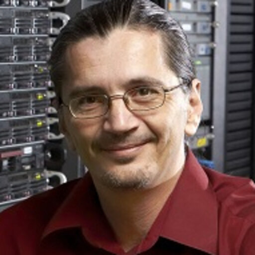
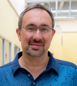
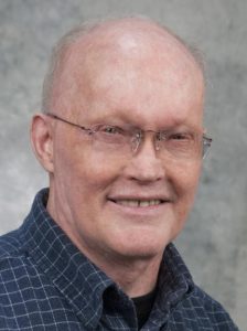
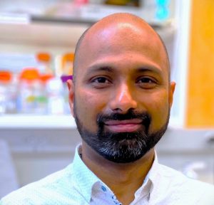
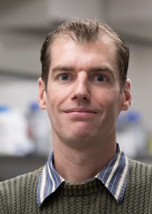
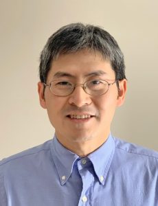
Pedro J. Mendes
|
CCAM Director Professor of Cell Biology Lab Website: Mendes Research Group. Faculty Directory
Research Interests: My research is in the area of computational systems biology, which aims to better understand biological systems through the use of computer models. My active areas of research include the development of modeling and simulation software as the author of the popular simulator Gepasi and leader of the COPASI simulator (with U. Kummer) and have been actively involved in the development of SBML, the systems biology markup language, and the MIRIAM proposal for model annotation. My group also constructs biochemical models, currently, this involves models of iron metabolism, eukaryotic translation, and microbial central metabolism. Through this work, I have pioneered the application of numerical global optimization in biochemical kinetic modeling and I am interested in using formal systems identification techniques in systems biology, particularly for reverse engineering models from data. My research requires a broad interdisciplinary approach and I work with people from most areas of science, either in my own research group or as collaborators.
Lab members: — Karen Osei-Boamah – MD/PhD student working on models of iron metabolism |
|
|---|---|---|
Ion Moraru
|
CCAM Deputy Director
Research Interests: My general research relates to the theoretical understanding of cellular processes, and developing tools for mathematical modeling in cell biology. Current work focuses on interfacing pathway/logic models and –omics data with kinetic quantitative models and dynamic simulations, both on new technology developments and the application to complex modeling of signaling pathways in normal and malignant cells. I am also the current lead developer of the Virtual Cell software platform for mathematical modeling and direct development of both the user interface and computational infrastructure. In addition, my interest in model reproducibility has led to a powerful new web platform that provides a central portal for simulating a broad range of models of biological systems. I have been involved in community standards for a long time, contributing to the development of both Systems Biology Markup Language (SBML) and Simulation Experiment Description Markup Language (SED-ML). |
|
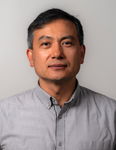
Yi Wu
|
Director of CCAM Microscopy User Facility
Research Interests: Research in our laboratory focuses on developing quantitative imaging tools that reveal dynamics of cellular signaling at high spatial and temporal resolution (biosensors), or that enable optical control of signaling proteins at with temporal and spatial precision (optogenetics). These tools are used with live cell microscopy to understand signaling networks underlying cellular function including cell polarity cell motility, axon guidance, and development of dendritic spines in neurons. In building biosensors, we use structural design strategies with fluorescent proteins to optimize FRET-based activity reporters for signaling proteins. Multiple live cell imaging modalities are used in the lab including TIRF, confocal, intensity-based ratiometric imaging, and fluorescence lifetime microscopy (FLIM). Optogenetics has generated much interest as it allows precise control of signaling in living systems using genetic engineering of natural photosensory proteins. We are exploring the use of the flavin-binding LOV (light-oxygen-voltage) domain from the plant photoreceptor phototropin. We used this technology to produce a genetically-encoded photoactivatable analog of Rac (PA-Rac) that enables precise modulation of Rac activity at regions that are submicrons in size and capable of controlling activation with microsecond precision in living cells. Current projects in the lab focus on extending such technology to other signaling proteins.
Lab members: — Milda Stanislauskas is a PhD student whose work explores how cells sense physical cues from the external environment and convert them into chemical signals. |
|
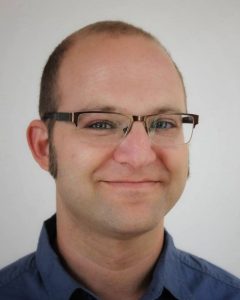
Eran Agmon
|
Assistant Professor of Molecular Biology and Biophysics
Research Interests: The Agmon lab builds comprehensive, multiscale models of cells—capturing everything from molecular mechanisms to population-level behaviors of diverse, interacting cells. With biological research now generating massive datasets on cell composition, dynamics, and spatial organization, we need new ways to turn this data into predictive, integrative simulations. We develop models that combine multi-source, multi-level data with diverse representations of biological function to decode data, generate hypotheses, and uncover organizing principles of cellular systems. We’re building Vivarium—an open-source platform for modular biological modeling—to support this vision. We are using it to develop the most complete whole-cell model of E. coli to date, simulate marine and gut microbiomes, explore tissue-level dynamics, and build foundational infrastructure and standards for the biological modeling community. |
|
Michael L. Blinov
|
Associate Professor of Genetics and Genome Sciences
Research Interests: My major research focus is on the computational modeling of signal transduction networks. Such networks often exhibit combinatorial complexity: the number of possible protein complexes and modification states is large. To accommodate this complexity we develop software to enable rule-based methods to specify a biological system using a set of rules. Using this rule-based approach, we create mathematical models of signal transduction networks in a single cell. We are specifically interested in describing activities and interactions among domains of biomolecules (e.g. phosphorylation of specific tyrosine residues, interactions between SH2 domain and phosphotyrosine) for receptor signaling and proteins-DNA interactions. My research is also focused in the bioinformatics arena, developing tools and methods for integrating public databases with mathematical modeling approaches. Data retrieval, visualization, flexible querying, and model annotation for future reuse, are some of the important requirements for modeling-based research in the modern age. In particular, we are developing ModelBricks as small, well-annotated, reusable model components that can be combined to develop complex models of biology. |
|
John Carson
|
Professor Emeritus of Molecular Biology and Biophysics
Research Interests: My laboratory investigates intracellular trafficking of mRNA in cells of the nervous system using a combination of molecular biological, microinjection, confocal microscopy, fluorescence correlation spectroscopy, and mathematical modeling techniques. We have delineated an RNA trafficking pathway consisting of export from the nucleus, granule assembly in the perikaryon, transport of RNA granules in the processes, localization to the distal regions of the cell, and translational activation. We are analyzing the cis/trans regulation of each step in this pathway and have identified a standard 21 nucleotide RNA trafficking signal (RTS) in many different transported mRNAs that bind specifically to hnRNPA2 and are involved at various steps in the trafficking pathway. |
|
Ann Cowan
|
Professor Emerita of Molecular Biology and Biophysics
Research Interests: I combine high-resolution quantitative imaging and biochemical analyses with mathematical modeling to develop computational simulations of specific cellular events using the Virtual Cell (VCell) modeling environment, a tool for creating spatially realistic mathematical models of cellular processes. I am also involved in the continued development of VCell and in developing a library of small, well-annotated models of specific cellular processes (ModelBricks) that can be used to build larger complex models of cellular events. As the first Director of the CCAM Microscopy User Facility, I established numerous collaborations based extensively on projects that apply quantitative fluorescence imaging to a wide range of cellular systems. |
|
Abhijit (Abhi) Deb Roy
|
Assistant Professor of Cell Biology
Research Interests: We are an experimental cell biology lab interested in both hypothesis-based and curiosity-driven questions, broadly within the realm of biomedical sciences. Our ongoing research projects are focused on investigating molecular signaling pathways and cytoskeletal dynamics involved in mechanobiology and cell migration. Directional cell migration plays critical roles during physiological processes such as development, angiogenesis and immune response, whereas dysregulation of cell migration is observed in pathologies such as cancer metastasis and atherosclerosis. We are interested in understanding how spatiotemporal regulation of signaling pathways modulates cytoskeletal dynamics to coordinate cell migration through diverse mechanical and biochemical environments. We employ live cell microscopy with biosensors and synthetic-biology based molecular actuators (optogenetic or chemical-inducible) to examine spatial and temporal relationships between molecular signaling pathways and cell behavior in a quantitative manner. We also collaborate with theoretical and computational biologists to develop predictive models bridging experimental observations to gain better understanding of complex biological systems. Ongoing projects: |
|
Michael Guertin |
Associate Professor of Genetics and Genome Sciences Guertin Lab. Faculty Directory
Research Interests: Precise regulation of transcription is central to cellular homeostasis and an organism’s response to developmental, nutritional, and environmental signals. Transcription factors bind to DNA to directly regulate gene expression and control cell fate. My lab develops and adopts molecular genomics approaches to study regulatory cascades initiated by hormone signaling. Our current work focuses on the basic mechanisms of estrogen signaling in breast cancer and how signaling cascades are disrupted upon drug treatment. We also use integrative genomics approaches to study adipogenesis. A long-term goal of my group is to identify transcription factors that are responsible for hormone-resistant phenotypes in disease states. Graduate students: |
|
Kshitiz
|
Assistant Professor of Biomedical Engineering Kshitiz Lab. Faculty Directory
Research Interests: Our lab’s objective is to understand a cell’s interaction with its neighbors and the microenvironment. With a particular focus on the tumor microenvironment, we aim to unravel the language cells employ to converse with each other, how the grammar and the content of the language adapt to the environment, and how cells behave in response. We use a gamut of of techniques, including live microscopy, bioinformatics, tissue engineering, nanofabrication, cell patterning, evolutionary biology, and genetics to answer fundamental questions in biology and medicine. Our current physiological focus involves understanding how new phenotypes emerge within cancer populations by their neighborly interactions; the evolutionary basis of metastatic tolerance; as well as cardiac maturation for disease modeling.
Lab members: –Yasir Suhail is a postdoctoral fellow working on the dependence of cancer metastasis on the surrounding stromal environment. –Visar Ajeti is a postdoctoral fellow. –Ashkan Novin is a Ph.D. student. |
|
Leslie M. Loew
|
Professor Emeritus of Cell BiologyBoard of Trustees Distinguished Professor
Research Interests: My lab is focused on the development of new experimental and computational technologies to help us understand the mechanisms underlying cell function. We have a longstanding effort aimed at developing and characterizing fluorescent probes of membrane potential. This effort is continuing, using organic chemical design and synthesis to develop better more sensitive voltage indicators. This work has led to a continual interest in the organization of signaling pathways along the cell membrane and within the cytoplasm. We are interested in the very general question of how the intricate spatial organization of molecules in cells is used to control cell function. This is especially true of neuronal cells and we are actively investigating the cell biology of dendritic spines in brain tissue. This motivated us to develop a very general computational modeling software platform called the “Virtual Cell”, in which we have created a framework for using computer simulation to explore cell biological mechanisms. The models are built naturally from experimental images of cellular and subcellular structures combined with biochemical and electrophysiological data. We have used the “Virtual Cell” system to explain the pattern of electrical and signaling activity in neuronal and non-neuronal cells. Most recently, we have developed new software called SpringSaLaD that can model molecular interactions at the sub-cellular scale. This allows us to explore how the shape of individual molecules control their interactions, leading ultimately to cell-level responses.
Lab members: –Kelvin Peterson is a medical student going for his MD. He is studying the kinetics and mechanisms membrane-associated reactions during cell signaling. |
|
Bruce J. Mayer
|
Professor and Vice-Chair of Genetics and Genome Sciences
Research Interests: Our group is interested in mechanisms of signal transduction. The ability of a cell to receive signals from the surrounding environment and respond to those signals appropriately is literally a matter of life and death. Whether a cell will proliferate, differentiate, or die, where it will adhere or migrate, virtually all aspects of its behavior depend on the ability to accurately interpret signals. Not only is signaling critical for normal development and the day-to-day function of an organism, but disregulated signaling underlies many human diseases such as cancer and autoimmune disorders. It is now appreciated that one of the central elements of the signaling machinery is the highly regulated and specific formation of protein-protein complexes. The fact that signaling relies on the binding of proteins to each other presents extraordinary opportunities: binding can be used as a means of identifying critical components of signaling pathways, and also provides the basis for strategies to inhibit those pathways in the laboratory or the clinic. We use a combination of biochemical and cell biological techniques to understand signaling pathways such as those that control cell proliferation and the organization of the cytoskeleton. We are also actively pursuing novel proteomic approaches to identify functionally important protein interactions and to characterize interactions on a global scale.
Lab members: –Kazuya Machida is an associate professor in residence in the lab developing tools to map SH2 domain binding site utilization in cells as a cancer biomarker. |
|
Vladimir Rodionov
|
Professor Emeritus of Cell Biology
Research Interests: Research in my laboratory is focused on molecular mechanisms of intracellular transport and the organization of microtubule cytoskeleton. Melanophores, pigment cells of lower vertebrates, are large cells that synchronously transport thousands of membrane-bounded pigment granules either rapidly to the cell center to form a tight aggregate or to disperse uniformly throughout the cytoplasm. During aggregation, granules move along microtubules using cytoplasmic dynein, but dispersion involves both initial microtubule-dependent transport to the periphery by Kinesin II and then slow diffusion-like movement along randomly arranged actin filaments. Transport is regulated by a Protein Kinase A (PKA) signaling cascade and thus provides a unique system for studying the role of the cytoskeleton in intracellular transport, mechanisms of switching between the two major transport systems, and regulation of activity of motor molecules by signal transduction mechanisms. We use molecular biology and biochemistry along with live cell fluorescence microscopy, photobleaching, photoactivation, and microinjection of motor-specific probes to understand the molecular mechanisms underlying the regulation of intracellular transport. |
|
Sarvenaz Sarabipour
|
Assistant Professor of Cell Biology
Research Interests. Our group is interested in understanding the mechanisms of signal transduction in cells and tissue. To do so, we combine techniques in multiple quantitative biological disciplines including biophysics, molecular biology, biochemistry and computational systems biology from single molecule measurements to multi-scale models of physiology and pathophysiology. We build multi-scale models of cells from their molecular interactions to their function in tissue to identify signal transduction mechanisms that contribute to cellular proliferation and migration in vascular and neuronal cells. The knowledge we gain is key in finding ways to modulate these critical signaling pathways for therapeutic potential for disease. |
|
Boris Slepchenko
|
Associate Professor of Cell Biology
Research Interests: My research has been focused on developing and testing numerical algorithms for applications in cell biology, particularly related to the Virtual Cell software project. I also work with collaborators to develop novel mathematical models for applications in cell biology, including calcium dynamics, nucleocytoplasmic protein transport, cell cycle, cell signaling, diffusion in crowded intracellular environment, actin polymerization kinetics, actin mechanics and cell motility, and stem cell population dynamics. |
|
Paola Vera-Licona
|
Assistant Professor of Cell Biology Lab Website: veraliconalab.org/. Faculty Directory
Research Interests: Our research is at the intersection of computational systems medicine and systems biology, mathematical biology, and bioinformatics. We work on the design, software development, and application of mathematical algorithms for the modeling, simulation, and control of biological systems. In molecular biology, the systems of our interest include gene regulatory networks and intracellular signaling networks where we aim to understand and control the cells’ intricate regulatory programs. We are focused on Cancer research (cancer reversion mechanisms and reversion of chemotherapy resistance) in breast cancer and leukemia.
|
|
Ping Yan
|
Assistant Professor of Cell Biology
Research Interests: My lab is focused on developing membrane potential probes to image neuronal and cardiac activities. We use synthetic organic chemistry, genetic techniques, optical spectroscopy, and microscopy to decode cells’ secrets. |
