Our Labs
Canalis Laboratory
Ernesto Canalis, MD
Director, Center for Skeletal Research
Co-Director, UConn Musculoskeletal Institute
Professor of Orthopedic Surgery and Medicine
Emily Denker
Research Assistant 1
Lauren Schilling
Research Assistant 3
Sara Stewart
Laboratory Assistant

The Canalis Laboratory is known for the discovery of skeletal growth factors and has pursued important investigations on the role of growth factors and their antagonists in skeletal function. This work was extended to studies on CCN proteins, including NOV and connective tissue growth factor, and these proteins can modulate the cellular activities of osteogenic signals. The laboratory has made seminal contributions to our understanding of the mechanisms of glucocorticoid action in bone in an effort to explain the pathogenesis of glucocorticoid-induced osteoporosis and correct the disease. The laboratory’s recent work has centered on factors determining cell fate, osteoblastogenesis, osteoclastogenesis, chondrogenesis, and cellular function. These investigations include studies on the role of Notch and nuclear factor of activated T cells (NFAT) in osteoblast, osteoclast, and chondrocyte cell differentiation in vivo and in vitro. We have developed genetically engineered mouse models of human disease, including models reproducing the pathogenic variants associated with Hajdu Cheney syndrome and Lehman syndrome. Recently, a mouse model of non-classical osteogenesis imperfecta was created. These mouse lines have allowed the study of mechanisms relevant to disease pathogenesis and the development of novel therapeutic avenues, including the use of antisense oligonucleotides. The laboratory has been funded continuously by the National Institutes of Health (NIH) since the 1980s, and in 1990 received a MERIT Award from the National Institute of Musculoskeletal and Skin Disorders (NIAMS). Currently, the laboratory is funded by NIAMS.
Kumbar Laboratory
Sangamesh G. Kumbar, Ph.D.
Professor of Orthopedic Surgery
Department of Biomedical Engineering
Materials Science and Engineering
Sama Abdulmalik, Ph.D.
Postdoctoral Fellow
Suranji Wijekoon, Ph.D.
Postdoctoral Fellow
Maria Soosai Lasar, Allen Zennifer, Ph.D.
Postdoctoral fellow
Rosalie Bordett
Graduate Assistant
Elifho Obopilwe
Graduate Assistant
Aditya Ruikar
Graduate Assistant
Sai Sadhananth Srinivasan
Graduate Assistant
Khadija Basiru Danazumi
Research Assistant
Laxmi Vobbineni
Health Research Program Student
Christopher Jesse Garcia
Medical Student

The Kumbar laboratory specializes in the fabrication and characterization of micro nanostructures, with a focus on semi-synthetic polymers for tissue engineering and drug delivery applications. Semi-synthetic polymers consisting of synthetic and natural-based materials integrate the advantageous mechanical properties of synthetic materials while preserving the inherent biological functions of natural materials. These novel materials are fabricated into micro nanostructures to promote enhanced tissue regeneration and controlled drug delivery systems. Current ongoing projects in the laboratory include:
Bone Regeneration: Engineered Cellulose Putty and Growth Factor Alternatives
The lab aims to utilize cellulose, the most abundant biopolymer, to serve as a mechanically competent platform for bone regeneration. The lab has been successful in creating a mechanically competent cellulosic scaffold platform to serve as a material for bone regeneration. The combination of natural polymers with micro and nano-scale features enhanced the regenerative abilities of the scaffolds both in vitro and in vivo. Efforts are also made to deliver growth factor alternative bioactive molecules to activate certain pathways to influence bone healing. Current investigations aim to improve the properties of these natural polymeric materials.
Neural Tissue Regeneration: Harnessing Electrical and Chemical Stimulation for Enhanced Recovery
The lab focuses on the development of conducting, degradable polymers and novel structured scaffolds for the regeneration of neural tissue. The slow regenerative nature of nerve tissue poses particular challenges to the clinical world as nerve injuries onset by pain, trauma, and/or degeneration rarely heal to suitable functional conditions. The lab develops, characterizes, and tests conducting polymeric scaffolds in conjunction with electrical and chemical stimulation techniques and stem-cell strategies to enhance the rate and efficacy of nerve tissue regeneration.
Soft Tissue Regeneration: Advancing Wound Healing and Minimizing Scarring
By recapitulating the natural environment of tissues through the use of materials, cells, and biochemical cues, it is possible to regenerate tendon tissue. Polymeric micro-nanostructures in combination with electrical and chemical stimuli as well as delivery cells and biological factors to improve healing and regeneration. Ongoing studies are focused on strategies to reduce scar and enhance functional outcomes.
Innovative Drug Delivery for Pain Management and Neuromuscular Disorders
The lab is pioneering drug delivery systems, using degradable polymeric micro/nanostructures for localized administration of diverse drugs. Current projects, funded for intervertebral disc treatment, extend to theranostic nanocarriers targeting conditions like stroke. A key focus is on developing targeted drug delivery for prolonged pain management, including a non-opioid therapeutic. Our approach involves identifying chemical modulators for α6β4 nicotinic acetylcholine receptors in dorsal root ganglia, aiming for enhanced pain relief compared to existing solutions.
Lorenzo Laboratory
Joseph Lorenzo, MD, Ph.D.
Director, Bone Biology Research
Professor of Orthopedic Surgery and Medicine
Sandra Jastrzebski
Research Associate 1
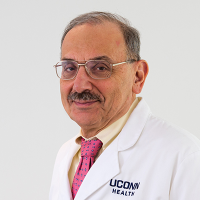
The Lorenzo Laboratory is known for its work investigating the interactions of bone and immune cells, a field now called osteoimmunology. The lab was among the first to examine how proinflammatory cytokines stimulated osteoclast-mediated bone resorption. Early papers demonstrated the mechanisms by which interleukin 1, tumor necrosis factor alpha and interleukin 6 stimulated an osteoclastic response. The lab also demonstrated that estrogen deficiency in animal models enhanced osteoclast-mediated bone loss, by mechanisms, which depended on the production of proinflammatory cytokines. After the discovery of the critical role that the protein receptor activator of nuclear factor kappa-Β ligand (RANKL) played in stimulating osteoclast formation and bone resorption, the lab demonstrated the reciprocal response relationship in bone between RANKL and its decoy receptor and RANKL-inhibitor osteoprotegerin (OPG). More recently the lab has done studies, which defined the phenotype of the osteoclast precursor cell and its relationship to other myeloid cells like macrophages and dendritic cells. Clinical and translational projects examined the role that the osteocyte product sclerostin had in bone mass differences in the production of RANKL between normal individuals and osteoporotic patients. Current projects are examining sexual dimorphisms in the rate that osteoclasts form and resorb bone, the role that the protein protease activated receptor 1 has in osteoclast function and differences in the phenotype and route of formation between osteoclasts that develop in homeostasis and those that develop with inflammation. The laboratory has been funded continuously by the National Institutes of Health (NIH), National Institute of Musculoskeletal and Skin Disorders (NIAMS) since 1981. In the past, additional funding has come from the Veterans Administration and industry.
Rux Laboratory
Danielle Rux, Ph.D.
Assistant Professor of Orthopedic Surgery

The Rux Laboratory specializes in musculoskeletal development, regeneration, and disease. Dr. Rux is an expert in skeletal developmental biology and in the Hox genes that are evolutionarily conserved for embryonic patterning. She received her Ph.D. in Cell and Developmental Biology from the University of Michigan, studying novel roles for the Hox genes in skeletal regeneration. Her postdoctoral research was done at the Children’s Hospital of Philadelphia, where her work led to novel insights into postnatal articular cartilage morphogenesis. She is the recipient of the NIH/NIAMS K99/R00 Pathway to Independence award.
Research in the Rux Lab is particularly focused on synovial joint development and articular cartilage morphogenesis. The joints are essential for a full range of motion and quality of life but are highly susceptible to diseases, including hip dysplasia and osteoarthritis. Unfortunately, synovial joint tissues have little to no capacity for innate regeneration, and damage is irreversible. A deeper understanding of the developmental processes that are vital to forming a fully functional joint will elucidate novel insights into disease and inform advanced strategies to prevent disease and/or boost regenerative capacity in patients. Dr. Rux’s highly innovative research program uses genetically modified mouse models combined with a range of techniques, including histologic imaging, single-cell and spatial transcriptomics, and in vitro culture, to elucidate morphogenetic mechanisms required for joint development and maintenance. Ongoing research projects center on defining how articular cartilage, the shock-absorbing cushion that lines opposing bones, acquires and maintains its zonal structure throughout life for optimal function. The lab is currently focused on patterning by the Hox genes and by the Hedgehog signaling pathway.
Sanjay Laboratory
Archana Sanjay, Ph.D.
Associate Professor of Orthopedic Surgery and Medicine
Chair, Biomedical Science Graduate Program
Justin King
M3 Graduate Student, Orthopedic Surgery and Medicine
Marta Stetsiv
M2 Graduate Student, Orthopedic Surgery and Medicine
Co-Mentored
Shagun Prabhu
Research Associate
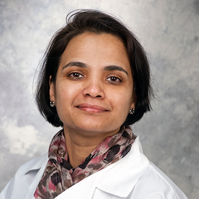
I have a broad background in bone research investigating bone formation and resorption processes during homeostatic and diseased states, including diabetes and fracture healing. Specifically, over the past few years, my NIH-funded study has been interested in examining the role of osteoprogenitors and signaling mechanisms that regulate bone healing response. My group has identified the Leucine-rich repeat-containing G protein-coupled receptor 6 (Lgr6) as a membrane receptor and adult stem cell marker essential for fracture healing. Another project in my lab focuses on aging and bone healing. Aging detrimentally impacts skeletal progenitors, reducing their quantity and differentiation abilities. Ongoing studies have shown that aged skeletal stem and progenitor cells exhibit reduced potential to enter the cell cycle and impaired osteogenic differentiation, affecting bone regeneration quality. Notably, the chemokine Cxcl9, linked to osteoporosis in men, was identified as significantly upregulated in osteoprogenitors derived from aged male mice. We are investigating sex-specific molecular pathways contributing to precision gerontology. This work was recently funded by the UConn Pepper Center’s Pilot and Exploratory Studies Core.
I have trained three postdoctoral fellows, eight graduate students, four dual-degree students, and several undergraduates. Graduate students and postdoctoral fellows who worked with me have received the highest-ranking career awards from national and international professional organizations and UConn Health.
I am the Chair of the Biomedical Graduate Program at UConn Health. The Ph.D. program in Biomedical Science at the UConn Health campus in Farmington, CT, emphasizes excellence in research training within an educational program in multiple disciplines.
I serve on study section panels for national and international organizations. I also serve as section editor for the Orthopedic Management of Fractures for Currents Osteoporosis Reports.
Sinder Laboratory
Benjamin Sinder, Ph.D.
Assistant Professor of Orthopedic Surgery
Renata Rydzik
Research Assistant 2
The Sinder Laboratory is focused on understanding skeletal growth and disease to develop new therapeutic strategies. Our laboratory is multidisciplinary and collaborative, with a combination of biological and biomechanical approaches. Specific areas of interest include the response of the skeleton to load, bone healing, bone biomechanics, and pediatric skeletal diseases. We are particularly interested in translating our basic preclinical research findings into improved patient outcomes. Dr. Sinder’s primary faculty appointment is in Orthopedic Surgery. He has a joint appointment in the Department of Biomedical Engineering, and he is also a faculty member of the Skeletal Biology and Regeneration graduate program at UConn Health.
UConn Spinal Research Laboratory
Isaac L. Moss, MD, CM, MASc, FRCSC
Professor of Orthopedic Surgery
Chair, Department of Orthopedic Surgery
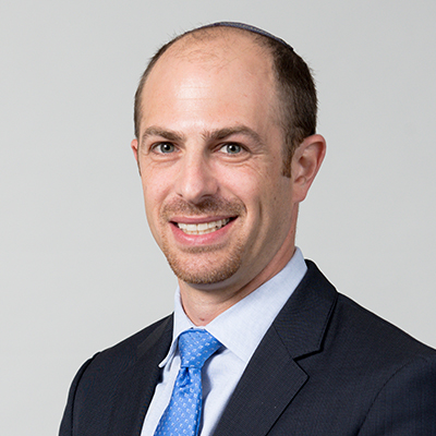
As a spinal surgeon, Dr. Moss has dedicated his career to the treatment of patients with spinal pathology to alleviate pain and improve function in a safe, efficient, and effective manner. Despite significant advancements in the field of spinal surgery, there is still room for improvement in the approach to many pathologic entities. Thus, Dr. Moss’s laboratory focuses its basic science research efforts on the development of biologics treatments to prevent and/or treat degeneration of the intervertebral disc in a minimally invasive and/or non-surgical manner. The lab employs both in vitro and in vivo preclinical models of disc degeneration to study pathology and evaluate novel treatments. Projects involve the utilization of advanced imaging techniques, such as atomic force microscopy, to gain a better understanding of the structural, biomechanical, and biochemical changes in the disc extracellular matrix associated with progressive degeneration. The lab also investigates the efficacy of growth factors, scaffold and cells for intervertebral disc regeneration. Specifically, they have demonstrated in both in vitro and in vivo studies that platelet-derived growth factor (PDGF) can slow the progression of degeneration by inhibiting disc cells apoptosis and promoting functional metabolism. Medical students, postgraduate fellows, and orthopedic residents all participate in research efforts.
