 Associate Professor
Department of Cell Biology
Contact
Phone: 860-679-2695
Email: terasaki@uchc.edu
markterasaki@me.com
Office: E6037
UConn Health
263 Farmington Avenue
Farmington, CT 06030
Research Interests
Structure and function of the endoplasmic reticulum; serial section electron microscopy; plasma membrane re-sealing; mechanism of nuclear envelope breakdown.
Selected Publications
Miki Furusho and Mark Terasaki. Evidence for glia-mediated, age-dependent remodeling of myelin at the axon initial segment. J Comp Neurol. 2024. Feb;532(2):e25574. doi: 10.1002/cne.25574.
Wood BM, Baena V, Huang H, Terasaki M, Kornberg TB. Cytonemes with complex geometries and composition extend to invaginations of target cells. J Cell Biol. 2021. 220(5):e202101116. doi: 10.1083/jcb.202101116
Ultrastructural Analysis of Cell-Cell Interactions in Drosophila Ovary. Methods Mol Biol. 2021. 2346:79-90. doi: 10.1007/7651_2020_342.
Analysis of the three dimensional structure of the kidney glomerulus capillary network. Sci Rep. 2020. 10(1):20334. doi: 10.1038/s41598-020-77211-x.
Odontoblast processes of the mouse incisor are plates oriented in the direction of growth. Anat Rec (Hoboken). 2021. 304 (8): 1820-1827. doi: 10.1002/ar.24570.
Baena, V., Owen, C.M., Uliasz, T.F., Lowther, K.M., Yee, S.-P., Terasaki, M., Egbert, J., Jaffe, L.A., Cellular heterogeneity of the LH receptor and its significance for cyclic GMP signaling in mouse preovulatory follicles. Endocrinology 2020. 161(7):bqaa074. doi: 10.1210/endocr/bqaa074.
2020. Ultrastructure of primary pacemaking cells in rabbit sino-atrial node cells indicates limited sarcoplasmic reticulum content. FACEB Bioadv. 2020. 2(2):106-115. doi: 10.1096/fba.2018-00079.
Extraocular, rod-like photoreceptors in a flatworm express xenopsin photopigment. eLife 2019. 8:e45465. doi: 10.7554/eLife.45465.
Baena V, Schalek RL, Lichtman JW, Terasaki M. Serial-section electron microscopy using automated tape-collecting ultramicrotome (ATUM). Methods Cell Biol. 2019;152:41-67.
Valentina Baena & Mark Terasaki. Three-dimensional organization of transzonal projections and other cytoplasmic extensions in the mouse ovarian follicle. Scientific Reports 9 (2019), Article number: 1262.
Bryan A. Niedenberger, Kenneth Cook, Valentina Baena, Nicholas D. Serra, Ellen K. Velte, Julio E. Agno, Karen A. Litwa, Mark Terasaki, Brian P. Hermann, Martin M. Matzuk, and Christopher B. Geyer. Dynamic cytoplasmic projections connect mammalianspermatogonia in vivo.
Terasaki M. Axonal endoplasmic recticulum is very narrow. J Cell Sci. 2018. 131(4): jcs210450.
Darras S, Fritzenwanker JH, Uhlinger KR, Farrelly E, Pani AM, Hurley IA, Norris RP, Osovitz M, Terasaki M, Wu M, Aronowicz J, Kirschner M, Gerhart JC, Lowe CJ. Anteroposterior axis patterning by early canonical Wnt signaling during hemichordate development PLoS Biol. 2018. 16(1):e2003698.
Yalçın B, Zhao L, Stofanko M, O’Sullivan NC, Kang ZH, Roost A, Thomas MR, Zaessinger S, Blard O, Patto AL, Sohail A, Baena V, Terasaki M, O’Kane CJ. Modeling of axonal endoplasmic reticulum network by spastic paraplegia proteins Elife. 2017. 6: e23882.
Norris RP, Baena V, Terasaki M. Localization of phosphorylated connexin 43 by serial section immunogold electron microscopy. J Cell Sci. 2017. 130: 1333-1340.
Terasaki, M. A finer look at a fine cellular meshwork. Science. 2016 Oct 28;354(6311):415-416.
Terasaki, M., Shemesh, T., Kasthuri, N., Klemm, R.W., Schalek, R., Hayworth, K.J., Hand, A.R., Yankova, M., Huber, G., Lichtman, J.W., Rapoport, T., and Kozlov, M.M. 2013. Stacked endoplasmic reticulum sheets are connected by helicoidal membrane motifs. Cell 154: 285-296. See also Cell Preview: Marshall, W.F. 2013. Differential geometry meets the cell. Cell 154: 265-266.
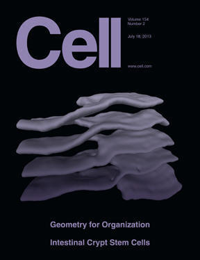
This paper explains how parallel sheets of endoplasmic reticulum are connected. The existence of these sheet stacks and their function in synthesis of membrane and secreted proteins has been known for more than 60 years. Three-dimensional reconstructions from serial section electron micrographs from the mouse salivary gland show that the sheets are connected by twisted membrane surfaces with helical edges. The overall structure resembles a parking garage, in which the different levels are connected by helicoidal ramps. Theoretical calculations show that this structure minimizes the elastic energy of sheet edges and surfaces and allows dense packing of this organelle in the restricted space of a cell.
Green, S.A., Norris, R.P., Terasaki, M., Lowe, C.J. 2013. FGF signaling induces mesoderm in the hemichordate Saccoglossus kowalevskii. Development 140: 1024-1033.
Darra, S., Gerhart, J., Terasaki, M., Kirschner, M., Lowe, C.J. 2011. b-Catenin specifies the endomesoderm and defines the posterior organizer of the hemichordate Saccoglossus kowalevskii. Development 138, 959-970.
Terasaki, M., and Runft, L. 2010. Two-stage dependence for 1-methyladenine induced reinitiation of meiotic maturation in starfish oocytes. Exp. Cell Res. 316: 2654-2663.
Lowe, C.J., Terasaki, M., Wu, M., Freeman, R., Runft, L., Kwan, K., Haigo, S., Aronowicz, J., Lander, E., Gruber, C., Kirschner, M., and Gerhart, J. 2006. Dorsoventral patterning in hemichordates: insights into early chordate evolution. PLOS 4: 1603-1619.
Terasaki, M. 2006. Fluorescence quantitation in thick specimens, with an application to cyclin B-GFP expression in starfish oocytes. Biol. Cell 98: 245-252.
Fein, A., and Terasaki, M. 2005. Rapid increase in plasma membrane chloride permeability during wound resealing in starfish oocytes. J. Gen. Physiol. 126: 151-159.
Lenart, P., Daigle, N., Bacher, C., Hand, A.R. , Eils, R., Terasaki, M., and Ellenberg, J. 2005. A contractile nuclear actin network drives chromosome congression in oocytes. Nature 436: 812-818.
Terasaki, M., Okumura, E., Hinkle, B., and Kishimoto, T. 2003. Localization and dynamics of Cdc2-cyclin B during meiotic reinitation in starfish oocytes. Mol. Biol. Cell 14:4685-4694.
Slepchenko, B.M., and Terasaki, M. 2003. Cyclin aggregation and robustness of bio-switching. Mol. Biol. Cell 14: 4695-4706.
Galbraith, J.A., and Terasaki, M. 2003. Controlled damage in thick specimens by multiphoton excitation. Mol. Biol. Cell 14: 1808-1817.
Hinkle, B., Slepchenko, B., Rolls, M.M., Walther, T.C., Stein, P.A., Mehlmann, L.M., Ellenberg, J., and Terasaki, M. 2002. Chromosomal association of Ran during meiotic and mitotic divisions. J. Cell Sci. 115: 4685-4693.
Terasaki, M., Runft, L., and Hand, A. 2001. Changes in organization of the endoplasmic reticulum during Xenopus oocyte maturation and egg activation. Mol. Biol. Cell 12: 1103-1116.
Terasaki, M., Campagnola, P., Rolls, M.M., Stein, P., Ellenberg, J., Hinkle, B., and Slepchenko, B. 2001. A new model for nuclear envelope breakdown. Mol. Biol. Cell 12: 503-510.