Syphilis/Treponema pallidum
 The syphilis spirochete T. pallidum (Tp) harbors many resemblances to B. burgdorferi but actually employs a markedly different parasitic strategy. Whereas B. burgdorferi is an enzootic pathogen, Tp is an obligate pathogen of humans which cannot be cultivated in artificial medium. The modes of transmission of the two bacteria differ markedly as well: Tp is transmitted from person-to-person during sexual activity, whereas Bb is transmitted by an arthropod vector. Once within the host, Tp begins to replicate locally, eventually causing a genital ulcer called a chancre, the clinical hallmark of the primary stage of the disease. As the chancre develops, treponemes begin to make their way towards draining lymph nodes and blood vessels in order to spread systemically. Once within the blood, Tp is extremely adept at invading virtually every organ system in the body, including the central nervous system, and establishing persistent infection that can cause serious, even life threatening, complications. We have designated Tp “the stealth pathogen” because of its remarkable ability to evade host immune defenses.
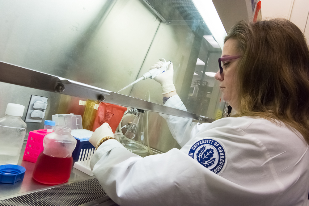
The clinical manifestations of venereal syphilis reflect the propensity of Tp to disseminate systemically and to induce chronic inflammation in diverse tissues and organ systems. The appearance of the syphilitic chancre during primary syphilis typically emerges 2 to 4 weeks after the initial contact with the spirochete. By this time, spirochetes have disseminated to other organs and a tissue, including the skin, setting the stage for what is classically known as secondary syphilis (SS), the focus of our translational syphilis research. Paradoxically, despite the robust nature of the adaptive cellular and humoral immune responses typical of SS, which includes the production high titers opsonic antibodies, it takes weeks and in some cases months for host defenses to gain control of the invading pathogen, ultimately giving rise to the prolonged asymptomatic stage known as latent syphilis. Efforts to understand the duality of immune evasion and immune recognition in syphilis have been hindered by the inability to propagate the bacterium in vitro and the lack of a suitable inbred animal model for performing immunologic studies. To circumvent these problems and obtain information directly relevant to the disease process in humans, we have been studying SS, the stage in which the dichotomous features of syphilitic infection are clearly evident and specimens are readily obtainable. Our clinical studies are complemented by our Tp structural biology program and a powerful ex vivo PBMC/monocyte-Tp stimulation model.
Our analysis of Tp’s unique ultrastructure provides potential explanations for the paradoxical features of secondary syphilis. Unlike the outer membrane of gram-negative bacteria, that of Tp lacks the potent proinflammatory glycolipid lipopolysaccharide (LPS). In addition, freeze fracture microscopy studies have shown that the spirochete contains very few outer membrane integral membrane proteins (OMPs). Although Tp does contain an abundance of highly antigenic hydrophilic polypeptides, these molecules are tethered by covalently bound N-terminal lipids to the bacterium’s periplasmic inner membrane leaflet. This unusual topology enables intact spirochetes to avoid sensing by innate immune receptors (i.e., Toll-like receptors) present on tissue macrophages and dendritic cells (DCs), while protecting the surface of the organism from the high titers of anti-treponemal antibodies that are generated during SS. Inefficient antibody binding to the sparse spirochetal OMP opsonic targets is thought to allow a large proportion of spirochetes to shun antibody binding and opsonophagocytosis; a mechanism which we have previously demonstrated is an essential requireme nt for Tp-driven innate immune cell activation. Fortunately for the host, a progressively more robust adaptive immune response, including enhanced uptake and degradation of opsonized spirochetes by tissue based macrophages and activation of CD4+ and CD8+ T-cells, eventually leads to control of bacterial replication and lesion resolution. Our ongoing translational studies are designed to elucidate the duality of immune evasion and immune recognition of the syphilis spirochete as it occurs in SS, and which builds upon recent advances from the Radolf laboratory in understanding the spirochete’s unique ultrastructure and composition and the host antibody responses it elicits in its natural human host.
Based on our combined observations, we are now able to propose a revised early syphilis pathogenesis model that integrates innate and adaptive immune responses to the bacterium and also takes into account the spirochete’s immunoevasive countermeasures against host defenses. According to this model, spirochetes replicate at the site of initial inoculation unchecked by the innate immune surveillance system and rapidly disseminate to the skin and other tissues. At some point after initial entry of the bacterium increasing local spirochetal burdens allow a small number of organisms to be taken up by resident phagocytes, although this process is inefficient in the absence of opsonic antibodies. APCs containing phagocytosed spirochetes can then migrate onto draining lymph nodes where they present treponemal antigens to naïve CD4+ T cells and B-cells. Neo-sensitized T-helper cells traffic back into the primary lesion, where they recognize their cognate antigens and release IFN-γ. Clearance of organisms by IFN-γ activated tissue macrophages is markedly facilitated by the emergence of high titers of Tp-specific opsonic antibodies. In parallel events, while the chancre resolves, as soon as treponemal loads in the skin of early syphilis patients reach a sufficient density capable of triggering the local inflammatory response, SS skin lesions become clinically apparent. In contrast to the immunologic events that initially take place in the primary chancres, innate and adaptive immune responses in SS skin lesions appear to co-evolve in the presence of primed CD4+ and CD8+ T cells and high titers of opsonic antibodies. One would thus predict that these changes would be sufficient for the immune response to eradicate the bacterium. However, the paucity of OMP antigenic targets on the outer leaflet of the bacterium together with the emergence of Tp-subpopulations resistant to opsonophagocytosis, permits varying numbers of bacteria to avoid opsonization and uptake by skin macrophages. The low-level bacteremia which ensues allows the spirochete to avoid recognition by host innate and adaptive immune defenses in the blood compartment. Chronic spread of Tp into other tissues, including the bone marrow, could affect the development of myeloid progenitors of monocytes, DCs and NK-cells. We hypothesize that the predominance of CD8+ T cells in SS skin lesions reflects a deviated cellular immune response, which could be prompted by less efficient priming of CD4+ T cells in lymph nodes. Fortunately for the host, over time, the emergence of greater numbers of antigen-specific CD4+ and CD8+ T-cells, IFN-γ producing CD56+ NK-cells together with increasing titers of Tp-specific opsonic antibodies, allows the host to ultimately gain the upper hand against the bacterium. The complex balance shift between immune evasion and bacterial persistence to immune recognition and spirochetal clearance, will thus not only determine the intensity and duration of the clinical manifestations of venereal syphilis but also how long the spirochete can endure in blood and tissues.
Lyme Disease/Borrelia burgdorferi
 Since Bb lacks orthologs of known exotoxins or the specialized secretory machinery required for the delivery of noxious molecules into host cells, it is widely accepted that the distinctive clinical signs and symptoms associated with Lyme disease result from the human host’s innate and adaptive immune responses to the bacterium. Monocytes and macrophages are considered to be two critical cellular elements of the innate immune response to Bb. Innate immune recognition of the spirochete by these cells was previously thought to result primarily from the interactions of the bacterium’s abundant outer membrane-associated lipoproteins with CD14 and Toll-like receptors (TLR) 1/2. However, experimental evidence generated in the Salazar laboratory has demonstrated that phagocytosed Bb induces inflammatory signals that differ both quantitatively and qualitatively from those generated by lipoproteins at the cell surface of monocytes/macrophages and dendritic cells. In our 2009 PLoS Pathogens publication (Salazar et al.), we demonstrated that internalized spirochetes induce transcription of interferon-β (IFN-β) and several type I interferon-stimulated genes (ISGs). In our 2011 PNAS publication (Cervantes et al.) we showed that recognition of borrelial ligands by monocytes involves a cooperative interaction between TLR2 and TLR8 signaling, and confirmed that endosomal TLR8 is solely responsible for IRF-7 mediated induction of IFN-β. More recently we proved that borrelial RNA is the principal TLR8 ligand and that in the case of Bb the TLR8-RNA interaction occurs solely in the phagosomal vacuole. Our combined observations have enabled us to formulate a new model for innate immune recognition of LD spirochetes. In this model, binding of unopsonized Bb to the monocyte/macrophage cell surface, through complement receptor 3 (CR3) (Hawley et al.) and other yet to be characterized phagocytic receptors, sets the stage for the broader immune signaling events that follow upon internalization of the bacterium and formation of the phagolysosome. As maturation of the phagosome proceeds and the spirochetes are degraded, TLR2 and TLR8 lining the vacuole sense spirochetal lipoproteins and borrelial nucleic acids, eliciting the production of pro- and anti-inflammatory cytokines, including TLR8-dependent induction of IFN-β.