Lyme Disease/Borrelia burgdorferi
 The Lyme disease spirochete B. burgdorferi (Bb) is maintained in nature via an enzootic cycle in which it is transmitted to a mammalian host (typically the white-footed mouse Peromyscus leucopus) by the nymphal stage of its arthropod vector, the deer tick Ixodes scapularis. The cycle is completed when the bacterium is acquired by a naïve larval tick feeding on an infected mouse. Infection of humans is not required for persistence of the spirochete in nature. In order to transit between its arthropod and mammalian hosts during the nymphal and larval blood meals, spirochetes must decipher complex environmental cues delivered at the feeding site and, in response, undergo dramatic changes in their transcriptomes and proteomes. The principal objective of Lyme disease research conducted in the Radolf Laboratory is to understand these processes. The alternative sigma factor RpoS is a unifying genetic feature of this project. Signals delivered by the nymphal blood meal induce the expression of RpoS. RpoS serves as a “gatekeeper” by transcribing genes required for transmission and establishing infection and by downregulating tick-phase genes no longer required once spirochetes reach their new mammalian host.
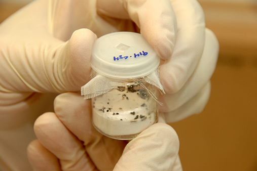
Most recently, we have begun dissecting other facets of the transcriptional and physiological adaptations Lyme disease spirochetes undergo during the enzootic cycle. One line of investigation, pioneered by Dr. Caimano, involves the two-component system (TCS) Hk1-Rrp1. This TCS is activated during the larval and nymphal blood meals and is not required for infection in mice. Without it, spirochetes are unable to survive within the noxious environment of the feeding tick midgut. The Hk1-Rrp1 brings about the adaptive changes required for survival during the blood meal by elaborating the signaling molecule cyclic-di-GMP (c-di-GMP). Production of c-di-GMP dramatically alters gene expression by Bb. One outcome is a global alteration of carbon utilization by the spirochete, enabling it to transition from the all-glucose environment of the mouse to the mixed carbon source environment of the tick midgut. Besides studying the numerous genes and genetic mechanisms activated by c-di-GMP, we also are elaborating the structure of the periplasmic domain of Hk1. This is the portion of the histidine kinase “environmental sensor” that binds exogenously produced molecules (as yet unidentified) that cross the outer membrane, bind to Hk1 within the periplasm, and initiate the signaling cascade that triggers the diguanylate cyclase activity of Rrp1, resulting in the synthesis of c-di-GMP.
Lyme disease spirochetes are extreme amino acid auxotrophs. They also have a limited repertoire of single amino acid transporters. How do these bacteria obtain the amino acids required for protein synthesis? We have found the answer. The spirochete uses an elaborate oligopeptide ABC transporter system to obtain peptide nutrients in both ticks and mice. The system relies on a repertoire of OppA oligopeptide binding proteins, one of which has been structurally characterized, to bind an assortment of peptides. Structural variations in these proteins, particularly their peptide binding cavities, along with variations in expression of individual OppAs during its enzootic cycle, enables the bacterium to precisely tailor its peptide acquisition system to the peptide ligands available in the environment.
Syphilis/Treponema pallidum
The syphilis spirochete T. pallidum (Tp) harbors many resemblances to Bb but employs a markedly different parasitic strategy. Whereas Bb is an enzootic pathogen, Tp is an obligate pathogen of humans which cannot be cultivated in artificial medium. The modes of transmission of the two bacteria differ markedly as well: Tp is transmitted from person-to-person during sexual activity, whereas Bb is transmitted by an arthropod vector. Once within the host, Tp begins to replicate locally, eventually causing a genital ulcer called a chancre, the clinical hallmark of the primary stage of the disease. As the chancre develops, treponemes begin to make their way towards draining lymph nodes and blood vessels in order to spread systemically. Once within the blood, Tp is extremely adept at invading virtually every organ system in the body, including the central nervous system, and establishing persistent infection that can cause serious, even life threatening, complications. We have designated Tp “the stealth pathogen” because of its remarkable ability to evade host immune defenses.
Efforts in the Radolf Laboratory to explain Tp’s stealth pathogenicity have focused on the bacterium’s unusual molecular architecture. Over the years, we have generated abundant evidence that the Tp outer membrane differs markedly in structure and composition from its Gram-negative counterparts (e.g., Escherichia coi). Not only does it lack lipopolysaccharide, the highly inflammatory glycolipid in the outer membranes of Gram negative bacteria, it also contains a much lower density of integral membrane proteins that present few surface antigenic targets to the host immune system. Situated below the outer bilayer, where they are inaccessible to antibodies in intact organisms, are the bacterium’s major immunogens, many of which are periplasmic proteins tethered by N-terminal lipids to the cytoplasmic membrane. This work ushered in what we have termed “the quest” for Tp outer membrane proteins, a project that has been ongoing for more than 20 years. Why a quest? Because identifying rare outer membrane proteins is so difficult and requires extraordinary commitment. In the past few years we have had great success using bioinformatics algorithms to identify outer membrane protein candidates based upon their predicted ability to form beta-barrels, the hallmark conformation of outer membrane proteins. Detailed topological and functional characterizations of a number of bona fide rare outer membranes identified using this strategy is now well underway.
Periodontal disease/Treponema denticola
The oral commensal spirochete T. denticola is a major component of the complex bacterial biofilm that causes periodontitis, a chronic inflammation of the tissues and bone that support the teeth. Our interest in T. denticola stems from (i) the need to better understand the microbiological factors that cause periodontitis and (ii) the ability to use this cultivatable, genetically manipulable organism as a surrogate for the noncultivatable T. pallidum. Our work with T. denticola centers around a protein called “the major outer sheath protein” or (MOSP), a known virulence determinant and porin, and the mechanisms for inserting newly synthesized outer membrane proteins such as MOSP into the T. denticola outer membrane. MOSP is closely related to a family of outer membrane proteins in T. pallidum. Thus, by studying its structure and function, we not only learn more about T. denticola host-pathogen interactions, we gain insights into an entire family of outer membrane proteins in the syphilis spirochete.
Leptospirosis/Leptospira interrogans
Leptospirosis, caused by multiple species within the genus Leptospira, is an emerging bacterial zoonotic disease with a worldwide distribution. Each year, >500,000 acute cases of leptospirosis are reported globally; when adjusted for potential under-reporting, this number increases to >1.7 million. Clinical manifestations of leptospirosis range from a mild febrile to a fulminant life-threatening illness. Case fatality rates for severe leptospirosis range between 10-70%. In poor urban communities in underdeveloped countries, major outbreaks of leptospirosis are associated with seasonal flooding.
L. interrogans is maintained in nature within a zoonotic cycle in which rats are the primary reservoir. By far, the most common route of infection in humans, who serve as incidental hosts, is through contact with water and soil contaminated with urine from reservoir hosts, primarily rats. Infection begins when a reservoir (or incidental) host comes into contact with contaminated water or soil. Once in the bloodstream, leptospires rapidly disseminate to multiple tissues. In reservoir hosts, leptospires are cleared within days from all sites except the kidney, where they colonize the proximal renal tubules. Infected reservoir hosts shed high numbers of leptospires in their urine for months to years. The ability of Leptospira to establish long-term residence within the kidney, a unique feature of leptospirosis, is a critical component of the bacterium’s pathogenic and zoonotic survival strategy. The development of novel strategies to reduce renal colonization in the reservoir host would have a direct impact on human disease.
Using a dialysis membrane chamber (DMC) rat peritoneal model, originally develop for B. burgdorferi, we performed the first RNAseq analysis of mammalian host adapted pathogenic Leptospira. These transcriptomic studies identified >160 genes that are differentially expressed by L. interrogans in response to mammalian host-specific stimuli, including genes is associated with physiological adaptation (i.e., nutrient uptake and utilization, redox homeostasis) as well as known virulence-determinants (e.g., collagenase, sphingomyelinases) and transcriptional regulators (e.g., a Fur/Per ortholog). The majority of genes upregulated in DMCs, however, encode hypothetical proteins of unknown function, making their contributions to virulence difficult to predict. The development of Transposon (Tn) mutant libraries for virulent L. interrogans now make it possible to functionally characterize differentially-expressed leptospiral genes and gene products involved in environmental sensing, renal colonization and persistence of leptospires within rats.