Multi-site recordings from the same dendritic branch reveal the role of dendritic voltage-gated Na channels in action potential backpropagation.
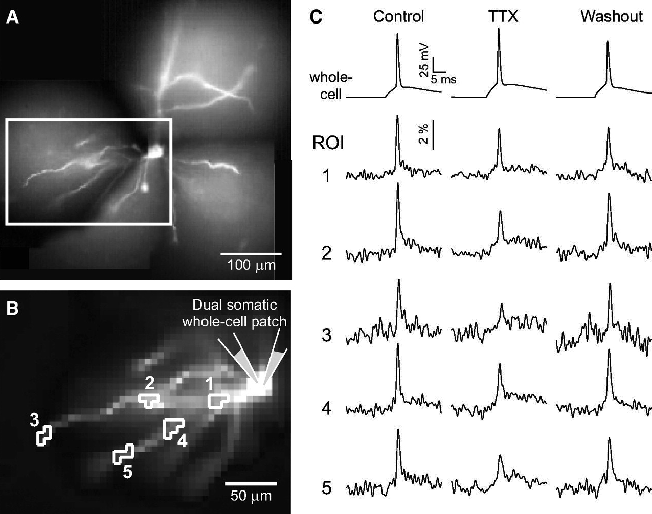
Figure 1. Multi-site voltage-sensitive dye imaging of backpropagating action potentials (bAPs) in basal dendrites of L5 cortical pyramidal neurons.
Dual somatic whole-cell patch. One patch pipette is in Voltage-Clamp Mode – it clamps the cell body in the shape of an action potential (AP playback). However, series resistance and capacitance prevent fast and accurate control of the cell body-voltage through voltage-clamp method. Therefore, it is necessary to compensate for the loss of AP amplitude (compensation factor). The second patch pipette is in Current-Clamp Mode – it allows us to determine the correct voltage-clamp compensation factor - it checks if the cell body is experiencing the correct AP waveform.
Dual somatic whole-cell patch is necessary in the TTX condition (Trial-2, middle column of traces). In all dendritic regions (e.g. 1-5), we compared voltage waveforms BEFORE and AFTER the complete block of dendritic voltage-gated Na channels (Fig. 1). We found the exact loss of AP amplitude when Na channels are blocked (Fig. 2).
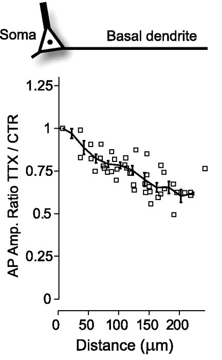
Figure 2. Contribution of Na channels to bAP amplitude in basal dendrites of L5 cortical pyramidal neurons.
In multiple recording sites along basal dendrites, we measured three parameters: [1] AP amplitude; [2] AP duration (half-width); and [3] AP latency. Each measurement was performed BEFORE and AFTER application of the Na channel blocker TTX. Based on these 6 values (three parameters, obtained before and after TTX), we constrained our multicompartmental model of L5 pyramidal cell (Fig. 3).
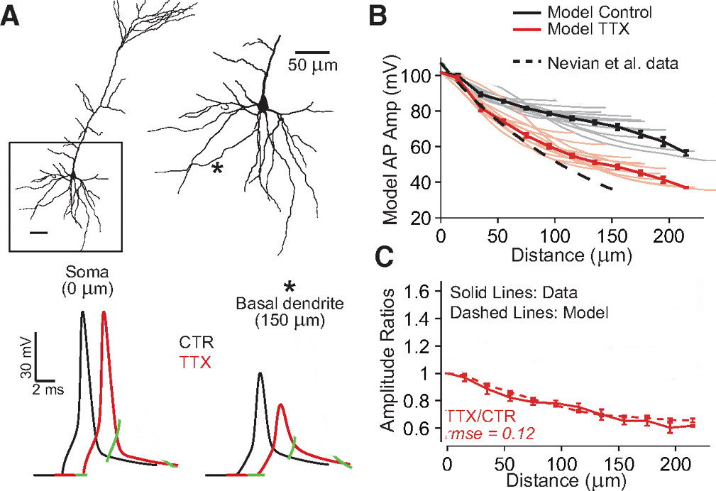
Figure 3. Best-fit model of action potential backpropagation in basal dendrites of L5 cortical pyramidal neurons.
In Fig. 3B, solid black line shows bAP amplitude along basal dendrites (on average). Solid gray lines represent individual dendritic branches in the model – there is some variability between branches. In summary, our experimental measurements (Figs. 1 and 2), combined with computational model (Fig. 3) reveal that bAP achieve much greater amplitudes in basal dendrites then those suggested by patch electrode recordings using very high-resistance patch electrodes (Nevian et al., 2007).
For precise spatial distributions of Voltage-gated Na channels and Voltage-gated A-type K channels in basal dendrites of L5 cortical pyramidal neurons see Acker and Antic, 2009.
Acker CD, Antic SD. Quantitative assessment of the distributions of membrane conductances involved in action potential backpropagation along basal dendrites. J Neurophysiol. 2009 Mar;101(3):1524-41.