Postdoctoral position in the laboratory of Dr. Srdjan D. Antic, Department of Neuroscience, University of Connecticut School of Medicine, Farmington, CT.
The research program is centered on three ongoing projects. Projects are not listed based on importance.
[Project A] Detecting physiological hallmarks of Alzheimer’s disease (AD) using mouse models of AD.
[Project B] Analysis of dendritic membrane excitability and dendritic integration using fast multi-site dendritic voltage imaging, whole-cell patch electrode recordings, glutamatergic stimulations, synaptic stimulations, optogenetic stimulations, and computational modeling. We are interested in several types of neurons in cortex and striatum.
[Project C] Developing and testing genetically-encoded calcium and voltage sensors for the analysis of cellular and circuit mechanisms in mouse cerebral cortex and striatum.
An ideal candidate should have experience with in vivo electrical recordings, in vivo optical imaging, acute brain slices, and intracellular electrophysiology. Some practical knowledge of programming (e.g., Matlab, Python) would be useful but not absolutely necessary. We cannot consider candidates who obtained a Ph.D. degree more than 10 years ago. Interested candidates should send a brief email and CV to Srdjan Antic.

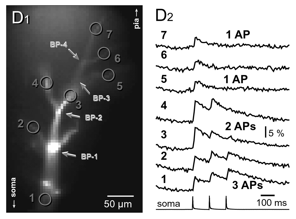
Filtering of action potentials in the apical tuft. D1) Microphotograph of a layer 5 pyramidal neuron in the rat prefrontal cortex. The neuron is filled with two fluorescent dyes (AlexaFluor 594 and Oregon Green Bapta-1). D2) Three action potentials were evoked by somatic current injections. Optical imaging of action potential-induced dendritic calcium transients was performed simultaneously from 7 locations (1 - 7) marked in D1. Bottom trace represents whole-cell recording from the cell body (soma). Three action potentials back propagate into the apical tuft dendrites and induce calcium transients. At each branch point (BP-2 to BP-4), one late AP is lost. Note that 3 spikes start from location number 1. The third spike is lost after passing through branch point BP-2. Two spikes start from location number 3. The second spike is lost after passing through branch point BP-4, so that only the first spike arrives in locations numbers 5, 6 and 7. Published in Neurophotonics.