Our Labs
Canalis Laboratory
Ernesto Canalis, MD
Director, Center for Skeletal Research
Professor of Orthopaedic Surgery and Medicine
Jungeun Yu
Instructor, Orthopaedic Surgery and Medicine
Magda Mocarska
Research Assistant 1
Lauren Schilling
Research Assistant 2
Sara Stewart
Laboratory Assistant

The Canalis Laboratory is known for the discovery of skeletal growth factors and has pursued important investigations on the role of growth factors and their antagonists in skeletal function. This work was extended to studies on CCN proteins, including Nov and Connective tissue growth factor, and these proteins can modulate the cellular activities of osteogenic signals. The laboratory has made seminal contributions to our understanding of the mechanisms of glucocorticoid action in bone in an effort to explain the pathogenesis of glucocorticoid-induced osteoporosis and correct the disease. The laboratory’s recent work has centered on factors determining cell fate, osteoblastogenesis, osteoclastogenesis, chondrogenesis, and cellular function. These investigations include studies on the role of Notch and Nuclear factor of activated T cells (NFAT) in osteoblast, osteoclast, and chondrocyte cell differentiation in vivo and in vitro. We have developed genetically engineered mouse models of human disease, including models reproducing the pathogenic variants associated with Hajdu Cheney Syndrome and Lehman Syndrome. Recently, a mouse model of non-classical osteogenesis imperfecta was created. These mouse lines have allowed the study of mechanisms relevant to disease pathogenesis and the development of novel therapeutic avenues, including the use of antisense oligonucleotides. The laboratory has been funded continuously by the National Institutes of Health (NIH) since the 1980s, and in 1990 received a MERIT Award from the National Institute of Musculoskeletal and Skin Disorders (NIAMS). Currently, the laboratory is funded by NIAMS.
Kumbar Laboratory
Sangamesh G. Kumbar, Ph.D.
Professor of Orthopaedic Surgery
Department of Biomedical Engineering
Materials Science and Engineering
Sama Abdulmalik, Ph.D.
Postdoctoral Fellow
Suranji Wijekoon, Ph.D.
Postdoctoral Fellow
Rosalie Bordett
Graduate Assistant
Elifho Obopilwe
Graduate Assistant
Aditya Ruikar
Graduate Assistant
Sai Sadhananth Srinivasan
Graduate Assistant

The Kumbar laboratory specializes in the fabrication and characterization of micro nanostructures, with a focus on semi-synthetic polymers for tissue engineering and drug delivery applications. Semi-synthetic polymers consisting of synthetic and natural-based materials integrate the advantageous mechanical properties of synthetic materials while preserving the inherent biological functions of natural materials. These novel materials are fabricated into micro nanostructures to promote enhanced tissue regeneration and controlled drug delivery systems. Current ongoing projects in the laboratory include:
Bone Regeneration
The lab aims to utilize cellulose, the most abundant biopolymer, to serve as a mechanically competent platform for bone regeneration. The lab has been successful in creating a mechanically competent cellulosic scaffold platform to serve as a material for bone regeneration. The combination of natural polymers with micro and nano-scale features enhanced the regenerative abilities of the scaffolds both in vitro and in vivo. Efforts are also made to deliver growth factor alternative bioactive molecules to activate certain pathways to influence bone healing. Current investigations aim to improve the properties of these natural polymeric materials.
Nerve Regeneration
The lab focuses on the development of conducting, degradable polymers and novel structured scaffolds for the regeneration of neural tissue. The slow regenerative nature of nerve tissue poses particular challenges to the clinical world as nerve injuries onset by pain, trauma, and degeneration rarely heal to suitable functional conditions. The lab develops, characterizes, and tests conducting polymeric scaffolds in conjunction with electrical and chemical stimulation techniques and stem-cell strategies to enhance the rate and efficacy of nerve tissue regeneration.
Tendon Regeneration
By recapitulating the natural environment of tissues through the use of materials, cells, and biochemical cues, it is possible to regenerate tendon tissue. Polymeric micro-nanostructures combined with electrical and chemical stimuli, delivery cells, and biological factors can improve healing and regeneration. Ongoing studies are focused on strategies to reduce scar and enhance functional outcomes.
Drug Delivery
The lab is developing multiple drug delivery systems using degradable polymeric micro/nanostructured vehicles. Injectable formulations can deliver a variety of drugs to treat various disease conditions in a local environment. Currently funded projects focus on the delivery of therapeutic agents in the intervertebral disc to halt disc degeneration. Ongoing work focuses on the development of theranostic nanocarriers that can deliver therapeutics to treat conditions like stroke. Targeted delivery of therapeutics for prolonged drug therapy systems and the treatment of cancer is of particular interest. A novel drug delivery platform seeking to mimic the tissue invasion strategy of the bacterium Listeria monocytogenes to treat cancers of various types has shown optimistic results. These drug delivery systems achieve monodispersed drug delivery in solid tumors to combat geometric drug resistance within certain regions of tumors that are distant from tumor vasculature and thus receive little to no drug.
Lorenzo Laboratory
Joseph Lorenzo, MD, Ph.D.
Director, Bone Biology Research
Professor of Orthopaedic Surgery and Medicine
Sandra Jastrzebski
Research Assistant 3
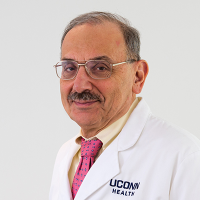
The Lorenzo Laboratory is known for its work investigating the interactions of bone and immune cells, a field now called osteoimmunology. The lab was among the first to examine how proinflammatory cytokines stimulated osteoclast-mediated bone resorption. Early papers demonstrated the mechanisms by which interleukin 1, tumor necrosis factor alpha, and interleukin 6 stimulated an osteoclastic response. The lab also demonstrated that estrogen deficiency in animal models enhanced osteoclast-mediated bone loss by mechanisms which depended on the production of proinflammatory cytokines. After the discovery of the critical role that the protein receptor activator of nuclear factor kappa-Β ligand (RANKL) played in stimulating osteoclast formation and bone resorption, the lab demonstrated the reciprocal response relationship in bone between RANKL and its decoy receptor and RANKL-inhibitor osteoprotegerin (OPG). More recently, the lab has done studies that defined the phenotype of the osteoclast precursor cell and its relationship to other myeloid cells like macrophages and dendritic cells. Clinical and translational projects examined the role that the osteocyte product sclerostin had in bone mass differences in the production of RANKL between normal individuals and osteoporotic patients. Current projects are examining sexual dimorphisms in the rate that osteoclasts form and resorb bone, the role that the protein protease activated receptor 1 has in osteoclast function, and differences in the phenotype and route of formation between osteoclasts that develop in homeostasis and those that develop with inflammation. The laboratory has been funded continuously by the National Institutes of Health (NIH) National Institute of Musculoskeletal and Skin Disorders (NIAMS) since 1981. In the past, additional funding has come from the Veterans Administration and industry.
Pilbeam Laboratory
Carol C. Pilbeam, MD, Ph.D.
Professor of Medicine and Orthopaedics
Associate Director, MD/Ph.D. Program
Emma Yocis, BA
Research Assistant

The Pilbeam lab was NIH funded for 25 years to study the regulation of bone resorption and formation with the ultimate goal of providing improved therapies for osteoporosis. Initial studies were on the role of prostaglandins (PGs) and their receptors in the physiology and pathology of bone. The lab was one of the first to publish on regulation in bone of the major enzyme responsible for PG production, inducible cyclooxygenase-2 (COX2). Our studies included the role of COX2 and PGs in osteosarcoma, bladder cancer, and the anabolic effects of hormones, growth factors, and mechanical loading. Recent studies identified a novel inhibitory factor of the anabolic effects of PTH. This factor, serum amyloid A3 (SAA3), was induced only in the presence of PGE2. It was produced by pre-osteoclasts and blocked the ability of PTH to induce cAMP in osteoblasts, thereby blocking osteoblastic differentiation. Deletion of Saa3 in mice changed the results of continuous PTH infusion from bone loss to bone gain, solving the longstanding conundrum of why intermittent but not continuous PTH is anabolic. We are now investigating the role of SAA3 in inflammation and its contribution to the chronic inflammation of aging. In addition, mentoring in the lab of students and junior faculty has been very important. Dr. Pilbeam has just stepped down from 10 years as the Director of our MD/Ph.D. program.
Sanjay Laboratory
Archana Sanjay, Ph.D.
Associate Professor of Orthopaedic Surgery and Medicine
Vice Chair, Biomedical Science Graduate Program
Justin King
M2 Graduate Student, Orthopaedic Surgery and Medicine
Matthew Wan
Research Assistant 1
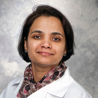
I have a broad background in bone research, investigating both bone formation and bone resorption processes during homeostatic and diseased states, including diabetes and fracture healing. Specifically, over the past few years, my NIH-funded research has been interested in examining the role of osteoprogenitors and signaling mechanisms that regulate bone healing response. Using an mRNA-Seq approach to identify novel transcripts that are upregulated upon injury, we found that expression of Lgr6, a G Protein coupled receptor in Wnt signaling, was significantly increased. Lgr proteins are adult stem cell markers in other tissues; however, the role of Lgr6 in bone healing and regeneration is undefined. Ongoing work in the lab has revealed that Lgr6 is required for robust bone healing response. Recently, we are also investigating the mRNA-micro- RNA networks in injured skeletal progenitor cells derived from intact and fractured bones in young, old, and diabetic mice.
I have trained two postdoctoral fellows, five Ph.D. students, two dual degree students, and several undergraduate students. All students trained in my laboratory have published consistently, and each has at least one first-author paper. Graduate students and postdoctoral fellows who have worked with me received the highest-ranking career awards from national and international professional organizations and UConn Health. A DMD/Ph.D. student trained in the laboratory also received the NIH F30 training award.
I serve as the Vice Chair of the Biomedical Graduate Program at UConn Health. The Ph.D. program in Biomedical Science at the UConn Health campus in Farmington, CT, emphasizes excellence in research training within an educational program in multiple disciplines. I recently served as Director of the Skeletal Biology and Regeneration Graduate Program at UConn Health. This field explores the cellular, molecular, and genetic processes related to skeletal development, skeletal diseases, injuries, and their regeneration.
UConn Spinal Research Laboratory
Isaac L. Moss, MD, CM, MASc, FRCSC
Associate Professor of Orthopaedic Surgery and Neurosurgery
Judy Kalinowski
Research Assistant
Nicolas Mancini
Medical Student Research Associate
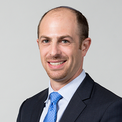
As a spinal surgeon, Dr. Moss has dedicated his career to the treatment of patients with spinal pathology to alleviate pain and improve function in a safe, efficient, and effective manner. Despite significant advancements in the field of spinal surgery, there is still room for improvement in the approach to many pathologic entities. Thus, Dr. Moss’s laboratory focuses its basic science research efforts on the development of biologics treatments to prevent and/or treat degeneration of the intervertebral disc in a minimally invasive and/or non-surgical manner. The lab employs both in vitro and in vivo preclinical models of disc degeneration to study pathology and evaluate novel treatments. Projects involve utilization advanced of advance imaging techniques, such as atomic force microscopy, to gain a better understanding of the structural, biomechanical and biochemical changes in the disc extracellular matrix associated with progressive degeneration. The lab also investigates the efficacy of growth factors, scaffold and cells for intervertebral disc regeneration. Specifically, they have demonstrated in both in vitro and in vivo studies that platelet derived growth factor (PDGF) can slow progression of degeneration by inhibiting disc cells apoptosis and promoting functional metabolism. Medical students, post-graduate fellows, and orthopaedic residents all participate in research efforts.
Youngstrom Laboratory
Daniel W. Youngstrom, Ph.D.
Director, X-Ray Microtomography (µCT) Core Facility
Assistant Professor of Orthopaedic Surgery
Jessica M. Kraus
Research Assistant 1
Renata Rydzik
Research Assistant 2
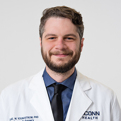
Bone is a dynamic tissue often capable of remarkable intrinsic healing, but there are limits to the extent of repair that is possible in humans. Yet some animals, including zebrafish, are enticingly capable of complete regeneration of complex skeletal structures following severe injury. What are the cellular mechanisms that give rise to this extraordinary healing potential? The Youngstrom Laboratory uses in vitro cell culture, pre-clinical animal models, and reverse genetics to discover ways to engineer adult skeletal stem cells and promote therapeutic bone formation following developmental malformation, surgical resection, or high-energy trauma. The current focus of the laboratory is Notch signaling during craniofacial regeneration in zebrafish.
